



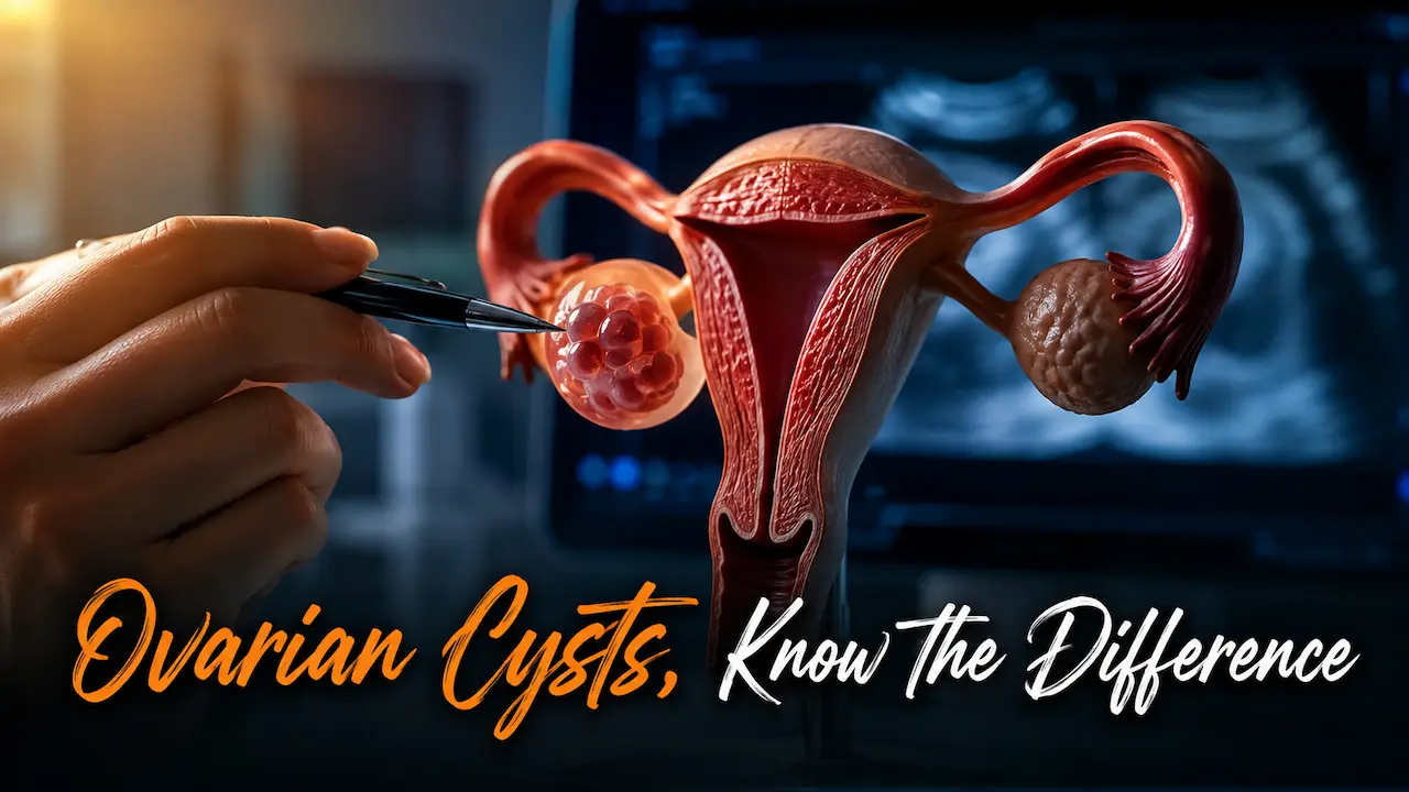
A scan report that says “cyst” rarely means the same thing twice. One patient’s cyst disappears in six weeks. Another’s needs surgery within the month. The difference almost always comes down to which of the types of ovarian cysts is actually showing up on the ultrasound, since size alone tells only part of the story.
Key takeaways
- Among the types of ovarian cysts, functional cysts are the most common and usually resolve without treatment.
- Dermoid cysts, cystadenomas and endometriomas are structural and generally need closer monitoring or removal.
- An endometrioma is a strong signal to also look into endometriosis, not just the cyst itself.
- The cyst type shapes the treatment plan more than size does, covered in full on the ovarian cyst treatment in Mohali page.
- A pelvic ultrasound remains the fastest way to tell one type from another.
What Doctors Mean When They Say “Type” of Cyst
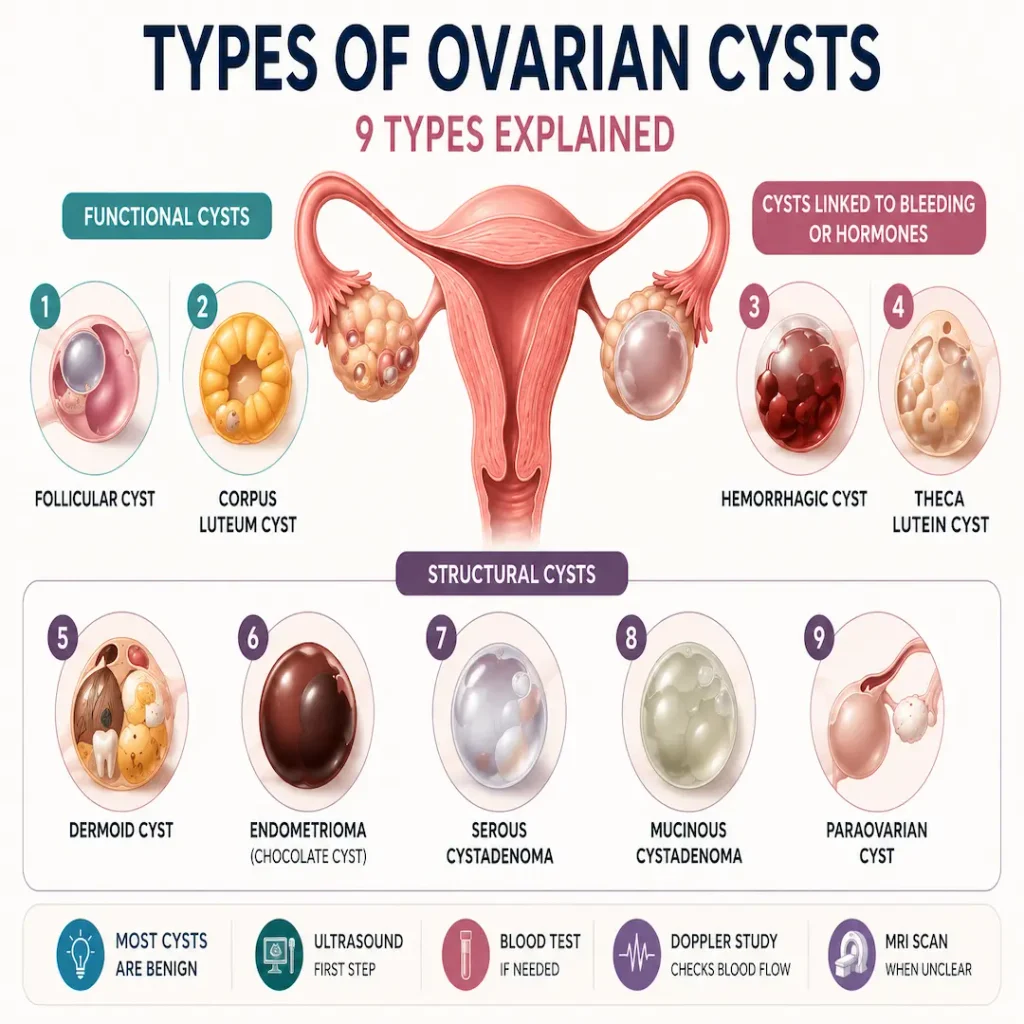
The types of ovarian cysts fall into two broad groups: functional cysts, which form as part of a normal menstrual cycle and usually resolve on their own, and structural or pathological cysts, which grow independently of ovulation and often need monitoring, medication or surgical removal.
That single distinction, functional versus structural, is what a gynecologist is really asking when a scan report uses an unfamiliar term. Each type below sits under one of those two branches.
Functional Cysts: The Two Common Types
A functional ovarian cyst forms directly from the process of ovulation, which is why these are by far the most common type found on a routine scan, according to NHS cyst guidance.
Follicular Cyst
Each month a follicle grows to release an egg. When it does not rupture on schedule, it can keep filling with fluid, forming a follicular cyst. Most sit under 3 centimeters, cause no symptoms and disappear within a cycle or two.
Corpus Luteum Cyst
Once an egg is released, the leftover follicle is called the corpus luteum. If it seals over and fills with fluid or blood instead of breaking down, a corpus luteum cyst forms. These can cause a dull ache on one side and occasionally delay the next period.
Cysts That Form From Bleeding or Extra Hormone
Some cysts are still linked to a normal cycle but behave differently once blood or extra hormone gets involved.
Hemorrhagic Cyst
When a follicular or corpus luteum cyst bleeds internally, it becomes a hemorrhagic cyst. Pain can arrive suddenly on one side, sharp enough at times to prompt an emergency visit, though most settle with rest and pain relief.
Theca Lutein Cyst
These form when ovarian tissue is exposed to unusually high hormone levels, most often during pregnancy or fertility treatment. They tend to appear on both ovaries at once and typically shrink once the hormone trigger settles.
Structural Cysts That Usually Need Monitoring or Surgery
Structural cysts grow independently of the menstrual cycle. They rarely disappear on their own, which is why this group gets more attention on a follow up scan.
Dermoid Cyst
Also called a mature cystic teratoma, a dermoid cyst is present from birth and can contain tissue such as hair, skin or teeth. Growth is slow, and most are found by accident. Removal is standard once confirmed, mainly to prevent later twisting of the ovary.
Endometrioma
An endometrioma forms when endometrial tissue attaches to the ovary and bleeds with each cycle, filling with old, dark blood that gives it the nickname chocolate cyst. Spotting one on a scan is often the first real clue that endometriosis is present elsewhere in the pelvis, not just on the ovary.
Serous Cystadenoma
A cystadenoma grows from cells on the outer surface of the ovary rather than from ovulation. The serous type is filled with thin, watery fluid, can grow noticeably large, and is generally removed once identified.
Mucinous Cystadenoma
The mucinous version is filled with thicker, gel like fluid and can grow larger than most other types before causing symptoms. Removal is usually recommended given how large these can get if left unmonitored.
Paraovarian Cyst
Technically not on the ovary at all, a paraovarian cyst forms in the tissue beside it, near the fallopian tube. It behaves like other simple fluid filled cysts, is often found by accident, and rarely needs treatment unless it grows large or becomes painful.
How Doctors Tell the Types of Ovarian Cysts Apart
Ultrasound remains the starting point for almost all cases.
- A transvaginal or pelvic ultrasound reads the fluid pattern, wall thickness and any solid components
- A CA125 blood test gets added when the scan looks complex or the patient is postmenopausal
- Doppler flow studies check whether the cyst has its own blood supply, a clue for structural types
- An MRI is used occasionally when the ultrasound picture still is not clear
According to Cleveland Clinic’s overview of ovarian cysts, most cysts are diagnosed this way without ever needing a biopsy, since imaging features alone usually point to a specific type. A full walk through of that process sits on our ovarian cyst diagnosis page.
Why the Type Changes What Happens Next
A follicular cyst under 3 centimeters and a mucinous cystadenoma of the same size get completely different plans, even though both would appear as “cyst” on a first read. Functional types usually mean a repeat scan in 8 to 12 weeks. Structural types more often mean a surgical consultation, since they will not resolve with time alone. The full breakdown of monitoring, medication and surgery by scenario is covered on the ovarian cyst treatment in Mohali page, and an endometrioma diagnosis specifically is worth reading alongside our ovarian cyst vs endometriosis comparison, since the two conditions are often managed together.
Ovarian Cyst Types Mohali and Chandigarh Patients Ask About Most
In practice, ovarian cyst types Mohali clinics report most often are the functional ones, simply because they get picked up on routine scans done for unrelated reasons. Dermoid cysts and endometriomas come up next, usually once a patient already has pelvic pain or a fertility concern driving the scan. Anyone reviewing a report locally, or comparing notes with an ovarian cyst specialist Chandigarh/Mohali clinics also refer patients to, will find the same nine categories used, since the classification does not change with geography.
Knowing the exact type on your report is the fastest way to know what to expect next. If your scan uses a term from this list and you want it explained against your own results, that conversation is worth having early rather than guessing. The types of ovarian cysts listed above are the same set any gynecologist will check your report against, so bring the scan itself to your next appointment rather than just the summary line.
Frequently Asked Questions
1. What is the most common type of ovarian cyst?
Among the types of ovarian cysts, follicular cysts are the most common by a clear margin, since they form during a completely normal step of ovulation. Most women develop at least one over their reproductive years without ever knowing it, because these cysts rarely cause symptoms and typically shrink on their own within a cycle or two. They are usually found only when a scan is done for an unrelated reason. Corpus luteum cysts are the second most common functional type. Together, these two functional types account for the large majority of cysts picked up on routine pelvic ultrasounds.
2. Which type of ovarian cyst is dangerous?
No cyst type is dangerous by default, but structural types such as dermoid cysts, cystadenomas and endometriomas carry a higher chance of needing surgery since they do not resolve on their own. Any cyst becomes a genuine concern if it twists the ovary, ruptures and bleeds internally, or shows solid or irregular features on scan. Postmenopausal women with a new cyst generally get evaluated more closely, since the risk profile shifts after menopause.
3. Can a dermoid cyst turn into cancer?
Dermoid cysts are almost always benign, and cancerous transformation is rare, occurring in a very small percentage of cases and typically in larger, longstanding cysts. That said, because dermoid cysts do not resolve on their own and can twist the ovary as they grow, most gynecologists recommend surgical removal once one is confirmed rather than long term monitoring. The surgery is usually straightforward and laparoscopic, removing the cyst while preserving healthy ovarian tissue wherever possible.
4. What does an endometrioma mean for fertility?
An endometrioma itself does not always affect fertility, but its presence usually signals endometriosis elsewhere in the pelvis, which can affect egg quality and tubal function over time. Larger endometriomas, or ones removed surgically, can also reduce ovarian reserve on the affected side. Anyone planning pregnancy with a known endometrioma should have a fertility focused conversation before deciding between monitoring and surgery, since the approach that best protects fertility is not always the same one that best treats the cyst.
5. How is a cystadenoma different from a functional cyst?
A functional cyst forms from ovulation and typically resolves within a few cycles without treatment. A cystadenoma grows from cells on the outer surface of the ovary, has nothing to do with ovulation, and does not go away with time. Cystadenomas can also grow considerably larger than functional cysts before causing symptoms, which is part of why they are usually removed once identified rather than monitored indefinitely, even in the absence of pain.
6. Do all ovarian cyst types need surgery?
No. Functional cysts, which make up most cases, are typically just monitored with a repeat scan and resolve without surgery. Structural types such as dermoid cysts, cystadenomas and larger endometriomas are more likely to need removal, mainly because they persist and keep growing. The decision depends on type, size and scan features together, not the word cyst alone, so two people with a same size cyst can end up with different plans.
7. Can ovarian cyst types change or turn into each other?
Not typically. A follicular cyst can become a hemorrhagic cyst if it bleeds, and that is the closest thing to one type shifting into another. Structural cysts like dermoids or cystadenomas do not start as functional cysts and then convert, they form independently from the beginning. What can happen is that a cyst initially read as simple on an early scan later shows features that reclassify it, which is one reason repeat imaging matters for anything that has not resolved as expected.
8. How big does an ovarian cyst need to be before it is worrying?
Size matters, but type matters more. A functional cyst under 5 centimeters is rarely concerning regardless of type, while a structural cyst of the same size may already warrant a surgical opinion because it will not resolve on its own. Cysts above 10 centimeters, or ones with solid or irregular components on scan, generally move toward closer evaluation no matter which category they fall into. Sudden pain, fever or vomiting with any cyst size needs same day medical attention.
9. What type of cyst causes the most pain?
Hemorrhagic cysts tend to cause the sharpest, most sudden pain, since internal bleeding stretches the ovary quickly. Large dermoid cysts and cystadenomas can cause a duller, more constant pressure simply due to their size, and either type can trigger severe sudden pain if the ovary twists around its own blood supply, a situation called torsion. Endometriomas often cause pain that builds gradually around periods rather than arriving suddenly, which is one clue that helps separate them from a bleeding functional cyst.
10. Are cystadenomas and cysts from PCOD the same thing?
No. PCOD involves numerous small, immature follicles across both ovaries caused by a hormonal imbalance, and these are not usually described as one of the standard cyst types at all. A cystadenoma is a single structural cyst that grows from surface cells of the ovary and has no connection to PCOD or ovulation patterns. The two get confused because both involve the ovary and both show up on ultrasound, but the appearance, cause and management are entirely different.